Archive for the ‘retina’ Category

Macular disease, cataracts and art

Thursday, October 24th, 2013

My wife and I are supporters of two art museums, one locally and the other in Denver. I also have a personal interest in eye problems, especially cataracts and macular disease, as my father had lost an eye as an intern (a paper cut led to an infection and, in those days, before antibiotics, there was concern about the other eye developing problems, a medical issue called sympathetic ophthalmia). In his late 80s he had a cataract in his remaining eye and, when he was examined by an ophthalmologist at the Cleveland Clinic, was found to have macular degeneration, a chronic eye disease  usually seen in people over 50.

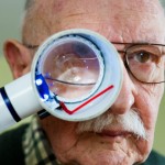
Someday my visions may deteriorate to this point.

Someday my visions may deteriorate to this point.

I became aware, as I read about Dad's problem, that one day it might become mine as well; one of the risk factors for macular degeneration is a family history of the disease.

I've pasted in a list of symptoms from this condition (copied from a Mayo Clinic website).

  • The need for brighter light when reading or doing close work
  • Increasing difficulty adapting to low light levels, such as when entering a dimly lit restaurant
  • Increasing blurriness of printed words
  • A decrease in the intensity or brightness of colors
  • Difficulty recognizing faces
  • A gradual increase in the haziness of your central or overall vision
  • Crooked central vision
  • A blurred or blind spot in the center of your field of vision
  • Hallucinations of geometric shapes or people, in case of advanced macular degeneration

The National Eye Institute, a branch of the NIH, has a fact sheet on age-related macular degeneration (AMD) that's worth looking at if you or someone in your family develops this problem. I'll mention a few things from that website as AMD is a major cause of vision loss in older adults. To begin with the macula is the part of your eyes that gives you the sharpest, most detailed vision. It's the extremely sensitive part of the retina, the layer of tissue  at the back of your eye that responds to light, converting images, focused by the eye's lens on this equivalent of camera film, into electrical signals that travel via the optic nerve to the brain. If the macula is damaged, fine points of these images become less clear.

If this happens to a non-artist, someone who doesn't make their living through images they put into a form that others can enjoy, it still leads to less sharp vision. You may have problems reading, driving or recognizing an image such as a face. Since your peripheral vision isn't affected, you'll probably be able to walk around without major difficulty.

But image that you're an artist. You gradually realize your vision is becoming less clear. You used to be able to read an eye chart at the 20/20 level, meaning you can read the same row of small letters on the chart at 20 feet which those with normal vision can. Now your visual acuity, measured when you see your eye specialist, is slipping and you worry that it will affect your ability to paint as well as you once did.

Having 20/20 eyesight does not necessarily mean perfect vision. 20/20 vision only indicates the sharpness or clarity of vision at a distance. There are other important vision skills, including peripheral awareness or side vision, eye coordination, depth perception, focusing ability and color vision that contribute to your overall visual ability.

Some people can see well at a distance, but are unable to bring nearer objects into focus. This condition can be caused by hyperopia (farsightedness) or presbyopia (loss of focusing ability). Others can see items that are close, but cannot see those far away. This condition may be caused by myopia (nearsightedness).

I've written about these medical problems before, but was riveted by a pair of articles I found in two AMA publications yesterday. A Stanford eye surgeon, Dr. Michael F. Marmor, just published a supurb article on Edgar Degas' progressive loss of vision in his later years. Degas was born in Paris in 1834 and died there in 1917. His painting altered from 1860 , when he had essentially normal vision, to 1870 and beyond  when first one eye, then the other progressively lost visual acuity. By 1897 he was seeing at a 20/200 level; that means he could would have to be twenty feet away from an eye chart to read the letters that someone with normal vision could read from 200 feet away.

The style and details of his paintings, especially his pastels, have been shown to change as Degas' eye problems progressed, but Dr. Marmor's article calls our attention to one oil painting, Scene from the Steeplechase: The Fallen Jockey. Here's a link to the painting in the National Gallery of Art; it was originally painted in 1866 and reworked by the artist in 1880-81 and again in 1896 with considerable changes made which Dr. Marmor shows can be linked to Degas' declining visual acuity.

A number of other significant artists have demonstrated visual loss in their work. An April, 2007 article in ScienceDaily focuses on Dr. Marmor's work, mentioning he's authored two books on art and eye sight: Degas Through His Own Eyes and The Artists's Eye (I've ordered a copy of the latter book through Amazon).

The Blind with Camera School of Photography website mentions a number of other famous figures from the art world who struggled with visual issues. Among those were El Greco, Rembrandt, Van Gogh, Paul Cezanne, Claude Monet, Mary Cassatt, Camille Pissarro and Auguste Renoir. Georgia O'Keeffe, who lived to the age of 98, also suffered with significant eye disease in her later years; her almost complete loss of eyesight and ill health during the last fifteen years of her life significantly curtailed her artistic productivity. Her eye problems began in 1968, and by 1971 macular degeneration caused her to lose all her central vision.

How is this honeybee similar to Monet?

How is this honeybee similar to Monet?

Monet had cataracts which not only diminished his visual acuity, but also affected his perception of colors. He resisted having surgery, but eventually decided to have one cataract removed. After the operation, according to science writer Carl Zimmer's review of the San Francisco Exploratorium's free  publication, Color Uncovered, Monet, like honeybees, was able to see ultraviolet light (normally filtered out by the lens of your eye) and painted water lilies a pale blue. Bees are guided to pollen by light signals we are unable to perceive; Monet had lost a lens to surgery, but gained a spectrum of light perception the rest of us lack.

I have zero talent as a visual artist, but after bilateral cataract surgery my vision is correctable to 20/20...for now.



The Eyes have it: Part two: diseases of the cornea and retina

Wednesday, June 19th, 2013

My own eye history is complex: I found out I was nearsighted at age eight while attending a baseball game with my folks. They mentioned the score and I asked how they knew what it was.

"Just read the scoreboard," Dad said.

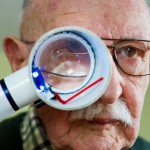
My eyesight isn't perfect, but I haven't gotten to this stage yet

My eyesight isn't perfect, but I haven't gotten to this stage yet

"What scoreboard?"

Much later, after years of wearing glasses (now they're bifocals), I was diagnosed as having a corneal disease...and that was even more years before the cataracts.

In my last post I outlined the anatomy and physiology of vision so that I could focus (pun intended) on diseases of the cornea and also on age-related macular degeneration this time.

In my prior post on the eye, I translated dystrophy as "it grew incorrectly," but "dys" comes from the Latin for "bad" and "trohe" from the Greek for "food," so poorly or inadequately nourished perhaps would be a better rendering for the term. A National Eye Institute document I provided a link to previously, "Facts about the Cornea and Corneal Diseases,"  says all of these dystrophies share a number of traits. They are usually inherited, affect both eyes equally, aren't caused by diet or injury, progress gradually and aren't related to systemic diseases, those that affect the rest of the body. They define a corneal dystrophy as "a condition in which one or more parts of the cornea lose their normal clarity due to a buildup of cloudy material."

Let's start with keratoconus, an abnormality of the cornea that usually affects both eyes, most frequently occurs in those who are ages 10 to 25 and slowly progresses over roughly a decade. It results in thinning and bulging of the cornea which forms a cone shape. This is a relatively uncommon disease affecting one in 2,000 of us and in its early stages can often be corrected with glasses or soft contact lenses.

Later on other kinds of contacts or even corneal transplantation may be required. If the patent gets such a transplant, its success rate is quite high with one estimate of an 89% positive outcome lasting ten or more years. An experimental treatment called collagen cross-linking is being evaluated around the world with clinical trials in multiple centers in the United States.

Less than 10% of those who have keratoconus also have a family history of the disease. Others may have had an eye injury, have other eye diseases, or unlike most other corneal problems, their keratoconus may be associated with systemic disorders including Down Syndrome.

My mother was said to have Fuchs' dystrophy, a slowly progressive corneal problem that normally affects both eyes, rarely alters vision until those who have it are in their 50s and 60s, and causes early-morning blurring that gradually clears as the day progresses. One layer of cells in the cornea, for unknown reasons, slowly dies off. They normally help pump water out of structure and, in their absence, the cornea swells, distorting vision.

Fuchs' is a major problem and some affected by this disease require advanced treatments including corneal transplantation. The success rate for this is reasonably good (73% after ten years), but waiting for donor tissue to become available is an issue.

I have map-dot-fingerprint (MDFD) dystrophy, according to most sources the most common of over 20 conditions in which parts of the cornea lose their usual clearness as cloudy materials pile up.

MDFD usually hits adults between the ages of 40 and 70, but can develop earlier. I remember cutting my right eye with a paper towel in 1971 when I was 30. It healed slowly and I had recurring episodes of eye pain, especially when I first woke up, over a period of four or five months. I'd bet that was my first hint of MDFD which typically will have flares over a few years and then go away without any long-last effect on vision.

A 2012 Medscape article says MDFD is rarely hereditary and may better be described as a corneal degeneration. Estimates of its prevalence in the general population range widely, from 2 to 43%. Like me, up to a third of those with MDFD have repeated early morning corneal tears/rips termed corneal erosions. They happen because in MDFD the outermost of the cornea's five layers of tissue, the epithelium, doesn't stay correctly attached to the layer below.

Since your eyes naturally get dry at night, your eyelid may stick to the epithelium and when you first awake and open your eyes, the not-well-attached epithelium can tear off casing mild to even severe pain.

I'm going to move to the back of the eye and age-related macular degeneration (AMD). Remember that light from images we see passes through the lens and goes to the retina, a light-sensitive layer of cells that line the inner surface of the eye. As the light impinges on these cells, two types of them react to the stimulus with a series of chemical/electrical events that eventually trigger nerve impulses sent through the optic nerve to the visual centers of the brain.  One kind is called rods and they are most important in dim light and provide our black and white vision. The other kind are termed cones and those are useful in our color perception and in daytime vision. I'll ignore a third type of receptor cells, important in reflexive responses to bright daylight.

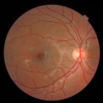
The macula is the dull round area slightly off-center

The macula is the dull round area slightly off-center

Near the center of a retina is a small, oval-shaped spot called the macula (Latin for "spot") that is critical for high-resolution vision. It's yellow in color, absorbs UV and blue light and is sometimes referred to as a natural sunblock protecting this highly-important area of the retina. The yellow color comes from two chemicals, lutein and zeaxanthin, which are derived from our diet. They are members of the carotenoids, a group of greater than 700 fat-soluble nutrients that provide the color in foods like carrots, pumpkins, sweet potatoes and other yellow, red, orange and deep green fruits and vegetables. Some of these, but by no means all, are converted to vitamin A or retinol, its active form, in the body.

AMD is the leading cause of major loss of vision in those of us over 50, accounting for more than half of all blindness in the United States. A major project, The Age-related Eye Disease Study (AREDS) has shown that taking some supplements can reduce the risk of progression to advanced AMD by 25% at 5 years. I've been taking a half dose of one of these for some years. Now a recent article in JAMA, "Lutein +Zeaxanthin and Omega-3 Fatty Acids for Age-Related Macular Degenerationpart of the followup study, AREDS2, looked at specific components of those formulations of antioxidant vitamins and minerals.

Their conclusion was adding the two carotenoids, lutein and zeaxanthin (L&Z), and the omega-3s  to the basic ingredients didn't seem to offer a further reduction in risk of AMD, but  L&Z might lower the risk of lung cancer in former smokers.

I'm not changing the pill I now take. I don't think ingesting the equivalent of a little extra carrot or sweet potato will hurt me.